
Paradigm Shift in Composite Restorations
The physical and chemical properties of composite resins have improved considerably in recent years and now offer, among other things, higher abrasion resistance (Spreafico and Roulet, 2009), improved biomimetic characteristics and, in particular, better control over polymerisation shrinkage. All these factors have resulted in a broader spectrum of indications for the use of composite resins (micro hybrid, nano filler and nanohybrid) in the posterior region. However, achieving an optimal approximal and occlusal anatomy as well as perfect restoration margins always remains a challenge, especially in the case of large cavities and hard-to-reach areas. In view of this, indirect partial restorations (e.g. onlays) are indicated in such clinical situations in which direct restorations are pushed to their technical limits. This is especially true in the case of complex cavities with margins in the direct vicinity of the gingiva or below the denlinoenamel junction.
The decision of whether a direct or indirect restoration is indicated is often a difficult one for dentists – especially since both options offer similar results with regard to their longevity (Van Dijken, 2000; Wassel et al., 2000; Pallesen and Qvist, 2003)
Whichever type of restorative treatment is ultimately selected, the objectives are always the same:
- Diagnosis and removal of carious lesions;
- Anatomical, functional and aesthetic restoration of the removed or absent dental tissue;
- Protection of pulp and dentine;
- Long-term preservation;
- Prevention of caries and periodontal recurrence
However, there are clinical situations, such as the loss of one or multiple cusps, approximal subgingival preparation margins and the preparation of approximal boxes with very open lateral walls that are far apart, in which the dentist is forced to turn to indirect techniques.
Why onlay?
Today, indirect restorations – in this case the onlay – are seen as the preferred form of treatment in the posterior region and a viable alternative to a full crown or a direct restoration. There are a number of associated factors:
- The result is on aesthetic and functionally well integrated, conservative restoration which allows both restorative (corrections prior to cementation and later repairs) and endodontic revision although this also applies for other restorations;
- Optimal occlusal anatomy and contact points without the complications sometimes associated with direct restorat
The problems associated with the direct technique resulted in the development of semi-direct techniques (Mormann et al. 1 9 83; Blonkenou et al. 1984; Mormann et al. 1989) with the aim of improving the quality of large Closs I and II restorations.
The disadvantages associated with onlays compared with direct restorations are:
- Dental laboratory costs and the associated time required – impact on cost for patient and patient compliance;
- Higher loss of healthy dental tissue associated with the build-up of divergent walls;
- At least two sessions required.
In light of the above, a semi-direct technique offers clear added value. It has some indications but offers a great advantage in that preparation, modelling and cementation of the restoration con be performed in a single session and at the same cost as a direct restoration.
An analogue technique is cost-effective, easy to perform and not associated with the high expense or problems involved with a digital chairside technique. As such, it can be seen as on additional option and ultimately as a rediscovered accomplishment of restorative dentistry.
The Semi-Direct Technique
This term exclusively applies to restorative techniques which involve both intraoral and extraoral steps and can be completed chairside in one treatment session. The restorations fabricated from composite are cemented using on adhesive technique.
Compared with intraoral restorations, extraorally fabricated restorations generally offer better anatomical and aesthetic potential, which is attributable to the more precise layering. This type of restoration is recommended in the following cases:
- Medium-sized cavities extending towards the dentinoenomel junction which rule out a direct technique or render it not recommend able;
- A limited number of teeth are affected.5
According to the literature, non-rigid models for onlays and onlays allow extraoral fabrication of restorations in a single visit procedure because these models cure quickly. The studies by Hirota R. et al. revealed that the predictability of the resell con be ensured by using the optimal combination of on alginate impression and a working model made of silicone, or a silicone or polyether impression and a working model mode of plaster.
The advantages of these techniques are illustrated in detail in the following case study.
Methods and Materials
Case study
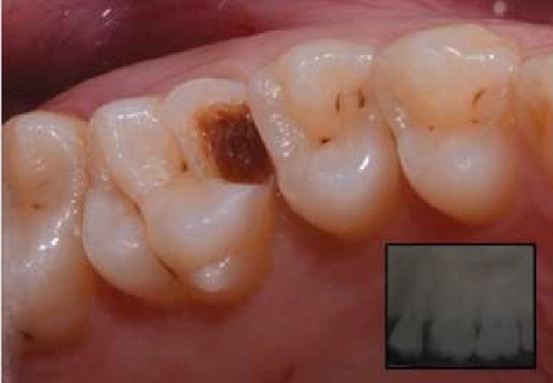
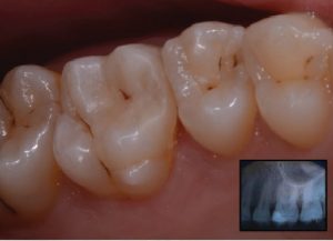
A 24 -year-old patient presented in the practice complaining of sensitively to cold in tooth 26 (upper left first molar) The
sensitivity was due to the loss of a composite restoration. According to the patient, the restoration had only been placed 11 months earlier using a single-visit procedure (Fig. 1).
With the exception of the morphofunctional deficiency of the tooth specified above, there was nothing remarkable in the patient’s medical or dental history.
Treatment
- An intraoral x-ray (Fig. 1, inset) was taken to exclude the possibility of endodontal involvement. We decided on restoration with an extraorally fabricated composite partial crown (semi-direct/indirect onlay) so as to ensure better predictability compared with a direct restoration, and also due to the absence of the mesiovestibular cusp, the thinness of the bevelled enamel and of the mesial margin of the cavity, which was close to the gingiva.
- Anaesthetic was applied in the area of teeth 26/27 and the operating site then isolated using a rubber dam (Fig. 2)
- The damaged dental tissue was removed and the cavity prepared in accordance with the adhesive guidelines.
- We performed “coronal repositioning of the margins” as described by Dietschi and Spreafico (1998)16 in order to simplify the clinical steps of the cementation procedure. This technique has proven its worth as an atraumatic alternative to clinical crown extension. It involves the placement of a matrix to ensure cervical sealing (in this case, a Tofflemire metal matrix), a 3-step etch -and -rinse procedure and cervical build-up with a flowable composite (x-tra base, VOCO Cuxhaven) with a maximal thickness of 1mm to reduce gingival micro leakage and improve marginal integrity.
Following successful build-up with a nanohybrid composite (Grar1dioSO, VOCO), we performed preparation of the tooth, comprising the mesiovestibular cusp and rounding off the preparation angle so as to remove undercuts, preserve as much dental tissue as possible and adapt the cavity walls (Fig. 3).
The prosthetic restoration can be fabricated using two different techniques:
- indirect technique in the dental laboratory;
- semi-direct chairside
We decided to fabricate one onlay with the first technique and another with the second technique so as to illustrate the advantages and disadvantages of each.
The indirect technique comprises
– Taking an impression with polyether (lmpregum, 3M) using an impression tray and a single-stage technique in both jaws (Werrin and Wilson, 1983);
Fabrication of a super-hard plaster model (type IV) with a model tray system and the opposing jaw;
Preparation of the plaster model for layering of the
composite (sectioning, application of the plaster hardening agent, blocking of the undercuts with wax and insertion of the anchorage);
Layering of the composite.
The semi-direct, extraoral technique (GrandioSO Inlay Syslem, VOCO) comprises:
Taking an impression with alginate in one jaw;
Drying the impression and fabrication of a model with addition-curing silicone (Die Silicone, VOCO);
Layering of the composite following complete curing of the silicone (4 mins)
The clear advantage of the chairside technique compared with the indirect technique is that no dental laboratory is required, which helps keep costs low. The significance of this aspect should not be underestimated, especially in patients with large carious lesions and where a prosthetic solution can be avoided for a long period of time without excessive expense.
When the working times for impression taking, model casting and fabrication (with the exception of the layering of the composite, as this is more or less comparable for both techniques) are compared, it becomes evident that the extraoral, semi -direct technique takes less time than the indirect (semi-direct = 5 minutes 45 seconds vs indirect = 1 hour 27 minutes).
The shortening of the working time makes it possible to fabricate the onlay in a single session. There is no need to insert a temporary restoration.
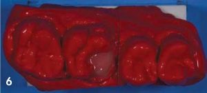
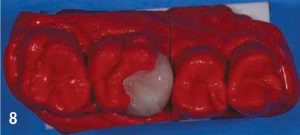
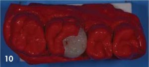
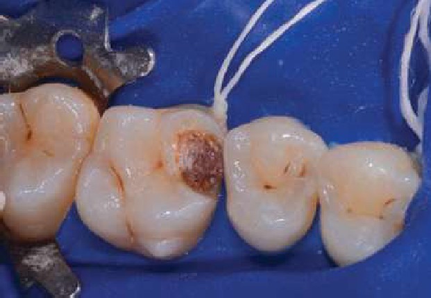
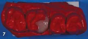
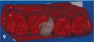
After making the model, we performed the layering of the composite (GrandioSO, VOCO) and fabricated two onlays for the same preparation (the individual steps for the layering of the composite on silicone are shown in figures 4 to 10).
Then an lwanson calliper gauge was used to compare the accuracy of fit of the silicone model cast in alginate with that of the super-hard plaster model cast in polyether. The width between two defined points (distal point of the preparation and the intersection point between the palatal gingival
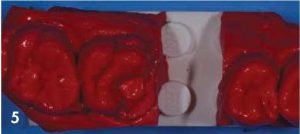
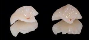
margin and the palatal intercuspal sulcus) was the same on both models (6 mm). The two onlays were switched on the models as an additional check of the accuracy of fit. No movement of the restoration and no marginal gap were observed.
The only disadvantage of the extraoral, semi-direct technique described in the literature is that the occlusal surfaces are built up without an opposing jaw model, and the requisite adaptations can therefore sometimes prove
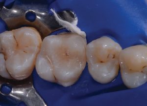
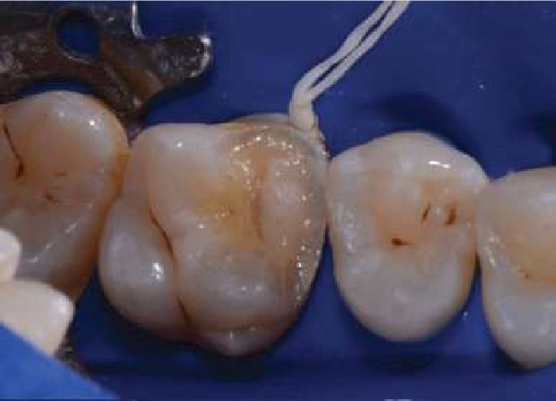
Following the layering, the restorations were finished, polished with diamond compound and sadnblasted with aluminium oxide/silcone dioxide. The surfaces were sealed with adhesive (Seal Coat, DEI, Italy)
We decided to cement the onlay layered on the silicone model. The cementation techniques comprises:
- Trying-in of the restoration;
- Isolation of the operating site;
- Cleaning of the tooth surfaces with chlorhexidine gel, pumice stone and a Robinson brush as well as sandblasting so as to produce efficient micro retention for luting cement;
- Selective enamel etching and application of dual curing self-etch bonding agent (Futurabond DC, VOCO) to the prepared area and the interior surface of the onlay;
- Injection of resin-based dual-curing cement (Bifix QM, VOCO) into the cavity;
- Placement of the onlay and removal of the occlusal excess using a probe and dental floss, application of glycerine gel along all margins and subsequent light curing for approx. 1.5 minutes on each side (Fig. 12).
This was followed by shaping and recontouring of the restoration using flexible polishing wheels with medium, fine and ultrafine grit sizes for the smooth approximal surfaces and with abrasive strips along the gingival margin. Any premature occlusal contacts were removed with a fine and ultrafine diamond bur.
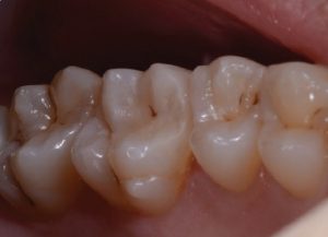
Figure 13 shows the finished clinical case after polishing with a single-stage diamond/silicone polisher (Dimanto, VOCO) and the perfect marginal integrity of the restoration following the intraoral follow-up radiograph.
The outstanding biomimetic integration of the restoration is still evident after 6 months (Fig. 14).
Conclusion
The extraoral, semi-direct technique has the same indications and advantages as the indirect technique, but additionally offers the convenience and the “single-visit advantage” of the direct chairside technique.
Considerable cost savings are also possible, as no laboratory or other technology is required. This technique heralded a paradigm shift in restorative dentistry.
References
- Hirata R. TIPS: dicas em odontologia estética. Brasil: Panamericana Publishing Co. Inc.,
- Brenna, Breschi, Cavalli, Devoto, Dondi, Dall’Orologio, Ferrari, Fiorini. Odontoiatria Restaurativa – Procedure di trattamento e prospettive Italy: Elsevier – Masson, 2009.
- Spreafico RC, Krejci I, Dietschi D. Clinical performance and marginal adaptation of class II direct and semidirect composite restorations over 3.5 years in vivo. J Dent July 2005; 33 (6): 499-507.
- van Dijken, Direct resin composite inlays/onlays: an 11-year follow-up. J Dent July 2000; 28(5): 299-306.
- Dietschi D, Spreafico R. Adhesive metal-free restorations: current concepts for the aesthetic treatment of posterior teeth. Berlin: Quintessence Publishing Co. Inc.,
- Gerrow JD, Price RB. Comparison of the surface detail reproduction of flexible die material J Prosthet Dent. Oct. 1998; 80 (4): 485-9.
- Price RB, Gerrow JD. Margin adaptation of indirect composite inlays fabricated on flexible dies. J Prosthet Dent. March 2000; 83(3): 306-13.
- Veneziani Restauri adesivi dei settori posteriori con margini cervicali subgengivali: nuova classificazione e approcio terapeutico differenziato. Il dentista moderno. Oct. 2008; 44-86.
- Magne P. Composite resins and bonded porcelain: the postamalgam era? J. Calif Dent Assoc. Feb. 2006; 34(2): 135-47.
- Kuroe T, Tachibana K, Tanino Y, Satoh N,Ohata N, Sano H, et al. Contraction stress of composite resin build-up procedures for pulpless molars. J Adhes Dent. Spring 2003; 5(1): 71-7.
- Mak YF, Lai SC, Cheung GS, Chan AW, Tay FR, Pashley Micro-tensile bond testing of resin cements to dentin and an indirect resin composite. Dent Mater. Dec. 2002, 18(8); 609-21.
- Swift EJ Jr, Perdigao J, Combe EC, Simpson CH, 3rd, Nunes Effect of restorative adhesive curing methods on dentin bond strengths. Am J Dent. June 2001; 14(3): 137-40.
- Van Meerbeek B, Perdigao J, Lambrechts P, Vanherle The clinical performance of adhesives. J Dent. Jan. 1998; 26(1): 1-20.
- Bayne SC, Heymann HO, Sturdevant JR, Wilder AD, Sluder TB. Contributing co-variables in clinical trials. Am J Oct. 1991; 4(5): 247-50.
- Kramer N, Lohbauer U, Frankenberger R, Adhesive luting of indirect restorations. Am J Dent 2000 13 (Special Edition): 600-760.
- Dietschi D, Spreafico R. Current clinical concepts for adhesive cementation of tooth-coloured posterior Pract Perio Aesthet Dent 1998; 10: 47-54.
- Estafan D, Estafan A. Flowable composite: a microleakage J Dent Res 1998; 77 (Special Edition B): 938-942.
- Labella R, Lambrechts, Van Merbeek B, Vanherle Polymerization shrinkage and elasticity of flowable composites and filled adhesives. Dent Mater 1999; 15: 128-137.
- Dietschi D, Olsburg S, Krejci I, Davidson C. In vitro evaluation of marginal and internal adaptation after occlusal stressing of indirect class II composite restorations with different resinous Eur J Oral Sci 2003; 111: 73-80.
- Chersoni S, Suppa P, Grandini S et al.In vivo and in vitro permeability of one-step self-etch J Dent Res 2004; 83(6): 459-464.
- Jiang W, Bo H, Yongchun G, LongXing N. Stress distribution in molars restored with inlays or onlays with or without endodontic treatment: a three-dimensional finite element J Prosthet Dent Jan. 2010; 103(1): 6-12.
- Shawkat E, Shortall A, Addison O, Palin Oxygen inhibition and incremental layer bond strengths of resin composites. Dent Mater 2009; 25: 1338-46.
- Park H, Lee I. Effect of glycerin on the surface hardness of composites after curing. JKACD 2011; 36 (6): 483-9.
- Dickinson GL, Leinfelder KF, Mazer RB, Russel CM. Effect of surface penetrating sealant on wear rate of posterior composite resins. J Am Dent Assoc 1990; 121: 251-255.
- Pettini F, Corsalini M, Savino M, Roselli G, Sibio G, Madeo D M, Pellegrino M, Di Venere D. Profilometric analysis of composite materials (microfilled, nanofilled and silorane) after different finishing and polishing procedures. Minerva Stomatologica 2014 – Volume 63, No. 4 Insert 1.


